Fleurettes Retinoblastoma : Retinoblastoma Dr Anupam Assistant Professor Ophthalmology Epidemiology The : 15% to 20% of retinoblastoma as harbor very well differentiated foci of actual photoreceptor differentiation.
Fleurettes Retinoblastoma : Retinoblastoma Dr Anupam Assistant Professor Ophthalmology Epidemiology The : 15% to 20% of retinoblastoma as harbor very well differentiated foci of actual photoreceptor differentiation.. Fleurettes are a higher form of photoreceptor differentiation than rosettes. Retinoblastoma is considered to be one of the types of embryonal central nervous system tumors, a group which includes medulloblastoma, neuroblastoma, pineoblastoma, and medulloepithelioma, among others. The degree of differentiation in retinoblastoma is determined by the development of rosettes and fleurettes, which has been confirmed by electron microscopy and tissue culture. 15% to 20% of retinoblastoma as harbor very well differentiated foci of actual photoreceptor differentiation. Fleurettes are considered the most differentiated form of rosette found in the tumor.
Necrosis is also very common and occurs when the tumor outgrows its vascular supply. Fleurettes are considered the most differentiated form of rosette found in the tumor. Traditionally, level of neoplastic differentiation is a crucial index of adjuvant chemotherapy and prognosis. 15,29 programmed cell death or apoptosis is also evident in the retinoblastoma. A benign variant of retinoblastoma termed retinocytoma or retinoma has been described.
Developmental Stage Specific Proliferation And Retinoblastoma Genesis In Rb Deficient Human But Not Mouse Cone Precursors Pnas from www.pnas.org Retinoblastoma protocol applies to retinoblastoma only. • appear years (median time 40 months) after successful treatment of. Incomplete shrinkage of the lesion may be noted after radiotherap). Necrotic cells appear pink on h&e staining. Only one patient died (20 years after enucleation) because of metastatic osteosarcoma.in conclusion: The degree of differentiation in retinoblastoma is determined by the development of rosettes and fleurettes, which has been confirmed by electron microscopy and tissue culture. Our results show that s antigen may be a useful marker in the study of the embryologic development of the human. • usually fatal due to meningeal spread, median survival of 9 months.
Fleurettes are retinoblastoma cells that have undergone greater photoreceptor differentiation.
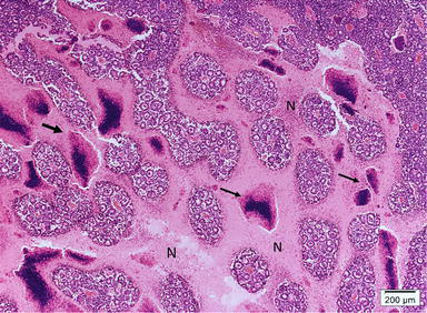
Fleurettes (figure 3c) are retinoblastoma cells that have undergone greater photoreceptor differentiation and group together as a bouquet. This comparative study showed that rosettes and fleurettes of retinoblastoma are an attempt to differentiate photoreceptor cells. Necrosis is also very common and occurs when the tumor outgrows its vascular supply. More rosettes, fleurettes & photoreceptor differentiation. January 2005 based on ajcc/uicc tnm, 6th edition procedures • cytology (no accompanying checklist) • biopsy (no accompanying checklist) • resection (globe) authors david l. • usually fatal due to meningeal spread, median survival of 9 months. • primary retinoblastoma of pineal & parasellar sites. Calcification is almost pathognomonic of retinoblastoma, but its etiology is not known. Retinoblastoma histopathology is a combination of undifferentiated cells and areas of tumor differentiation shown as rosettes and fleurettes. 15,29 programmed cell death or apoptosis is also evident in the retinoblastoma. The degree of differentiation in retinoblastoma is determined by the development of rosettes and fleurettes, which has been confirmed by electron microscopy and tissue culture. No other retinal cell types were found. Incomplete shrinkage of the lesion may be noted after radiotherap).
Fleurettes (figure 3c) are retinoblastoma cells that have undergone greater photoreceptor differentiation and group together as a bouquet. Pathology of retinoma reveals nonproliferative fleurettes. • usually fatal due to meningeal spread, median survival of 9 months. Calcification is common in these tumors. January 2005 based on ajcc/uicc tnm, 6th edition procedures • cytology (no accompanying checklist) • biopsy (no accompanying checklist) • resection (globe) authors david l.
Histopathological Characteristics And Classification For Prognostic Indicators Intechopen from www.intechopen.com 10 many retinoblastomas have underlying elements of retinoma. This comparative study showed that rosettes and fleurettes of retinoblastoma are an attempt to differentiate photoreceptor cells. Page, md department of pathology, vanderbilt university medical center, nashville. A tumor composed of fleurettes is deemed benign and called retinoma or retinocytoma. Fleurettes are a higher form of photoreceptor differentiation than rosettes. Tumors composed entirely of fleurettes (retinoma/retinocytoma) are thought to be retinoblastoma precursors, and like retinoblastoma, harbor mutations in both copies of the rb1 gene. Retinoma discovered in a retinoblastoma eye removed after the main active tumor dispersed throughout the vitreous following 1 cycle of chemotherapy. Retinoblastoma is a rare cancer of the infant retina that is diagnosed in approximately 8,000 children each year worldwide.
• appear years (median time 40 months) after successful treatment of.
Page, md department of pathology, vanderbilt university medical center, nashville. Only one patient died (20 years after enucleation) because of metastatic osteosarcoma.in conclusion: Retinoblastoma histopathology is a combination of undifferentiated cells and areas of tumor differentiation shown as rosettes and fleurettes. No other retinal cell types were found. 10,31 comparison of adjacent normal retina, retinoma, and retinoblastoma showed loss of both rb1 alleles and early genomic copy number changes in retinoma that were amplified further in the adjacent retinoblastoma. Incomplete shrinkage of the lesion may be noted after radiotherap). Fleurettes lack mitosis or necrosis. • appear years (median time 40 months) after successful treatment of. A tumor composed of fleurettes is deemed benign and called retinoma or retinocytoma. Necrotic cells appear pink on h&e staining. Fleurettes are considered the most differentiated form of rosette found in the tumor. This comparative study showed that rosettes and fleurettes of retinoblastoma are an attempt to differentiate photoreceptor cells. Need mutations in both alleles to inactivate rb gene, a.
15,29 programmed cell death or apoptosis is also evident in the retinoblastoma. Necrotic cells appear pink on h&e staining. Calcification is almost pathognomonic of retinoblastoma, but its etiology is not known. Pathology of retinoma reveals nonproliferative fleurettes. Retinoblastoma protocol applies to retinoblastoma only.
Pdf Correlation Of Ocular Ultrasonography With Histopathologic Findings In Intraocular Retinoblastoma Semantic Scholar from d3i71xaburhd42.cloudfront.net Traditionally, level of neoplastic differentiation is a crucial index of adjuvant chemotherapy and prognosis. Necrosis is also very common and occurs when the tumor outgrows its vascular supply. Retinoma histology showing abundant fleurettes and sparse cells with eosinophilic cytoplasm. A benign variant of retinoblastoma termed retinocytoma or retinoma has been described. Incomplete shrinkage of the lesion may be noted after radiotherap). Retinoma discovered in a retinoblastoma eye removed after the main active tumor dispersed throughout the vitreous following 1 cycle of chemotherapy. May be congenital but not recognized until ages 6 months to 2 years. 15,29 programmed cell death or apoptosis is also evident in the retinoblastoma.
American ophthalmology society first adopted the term retinoblastoma in 1926.
More rosettes, fleurettes & photoreceptor differentiation. It forms when both retinoblastoma gene (rb1) alleles are mutated in a. The degree of differentiation in retinoblastoma is determined by the development of rosettes and fleurettes, which has been confirmed by electron microscopy and tissue culture. Fleurettes lack mitosis or necrosis. True spontaneous regression of retinoblastoma is rare, but is probably due to extensive tumor necrosis and central retinal artery occlusion, resulting in phthisis bulbi. Incomplete shrinkage of the lesion may be noted after radiotherap). Necrotic cells appear pink on h&e staining. Fleurettes are considered the most differentiated form of rosette found in the tumor. January 2005 based on ajcc/uicc tnm, 6th edition procedures • cytology (no accompanying checklist) • biopsy (no accompanying checklist) • resection (globe) authors david l. Retinoblastoma is a major cancer treatment success story in developed countries where most deaths are caused by secondary tumors in germline mutation carriers. Fleurettes are retinoblastoma cells that have undergone greater photoreceptor differentiation. 10 many retinoblastomas have underlying elements of retinoma. Retinoblastoma histopathology is a combination of undifferentiated cells and areas of tumor differentiation shown as rosettes and fleurettes.
Page, md department of pathology, vanderbilt university medical center, nashville fleuret. All of the differentiated neoplastic cells were either photoreceptors or müller's cells.


0 Komentar